


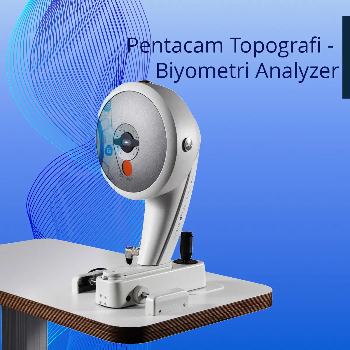
Pentacam Topography - Biometry Analyzer
What Is It Used For?
Pentacam is an advanced diagnostic device that captures and analyzes detailed three-dimensional images of the anterior segment of the eye — including the cornea, iris, lens, and anterior chamber. By generating a comprehensive map of the corneal structure, it supports the diagnosis of eye conditions, treatment planning, and surgical evaluation. It is used in the diagnosis of keratoconus and in assessing suitability for laser eye surgery or intraocular lens (IOL) implantation.
Advantage:
By producing a detailed map of the cornea — the eye's outermost transparent layer — Pentacam enables the creation of a personalized treatment plan tailored to each patient's unique anatomy.
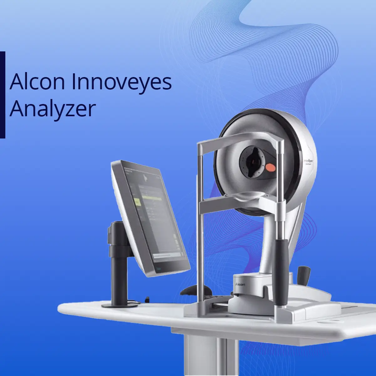
Alcon Innoveyes Analyzer
What Is It Used For?
It is used to select the appropriate intraocular lens (IOL) for cataract surgery. In refractive surgery, it provides a detailed assessment of the eye's optical structure and helps determine the most suitable treatment approach.
Advantage:
Precise biometric measurements simplify IOL selection for cataract surgery. It is an advanced diagnostic tool used to support accurate surgical planning and improve outcomes in eye surgery.
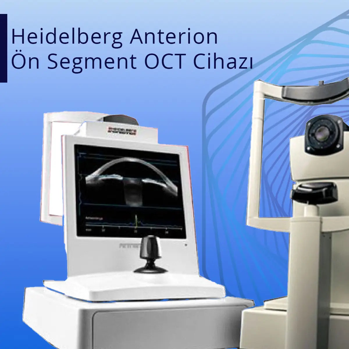
Heidelberg Anterion Anterior Segment OCT Device
What Is It Used For?
This device examines the anterior segment of the eye (cornea, iris, and lens), producing detailed images of intraocular structures.
Advantage:
Using OCT technology, it provides precise, non-invasive analysis of intraocular structures.
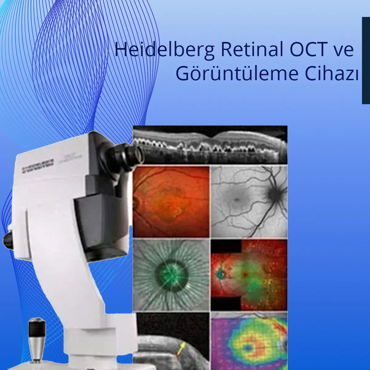
Heidelberg Retinal OCT and Imaging Device
What Is It Used For?
Provides detailed imaging of the retina and optic nerve. Used in the diagnosis and monitoring of retinal and optic nerve diseases.
Advantage:
High-resolution images allow rapid detection of problems in the retina and optic nerve.
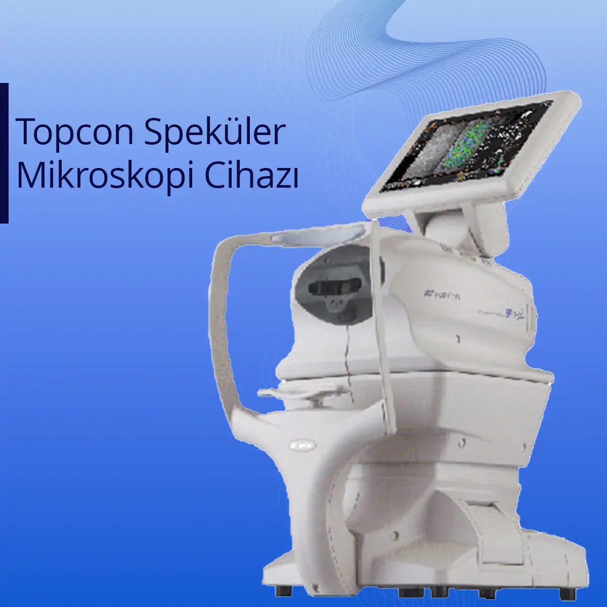
Topcon Specular Microscopy What Is It Used For?
What Is It Used For?
Specular microscopy is an imaging technique used to evaluate the endothelial cell layer on the inner surface of the cornea — the outermost layer of the eye. By assessing the number, size, and shape of endothelial cells, it provides detailed information about corneal health.
Advantage:
Specular microscopy is an important tool in assessing surgical risk, and serves as a valuable source of information both for diagnosis and for monitoring the course of treatment.
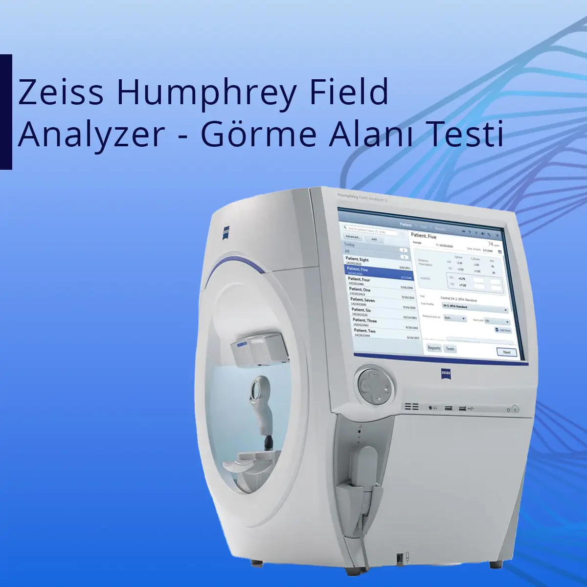
Zeiss Humphrey Field Analyzer – Visual Field Testing
What Is It Used For?
Glaucoma Diagnosis and Monitoring: Identifying narrowing and loss within the visual field is essential for diagnosing glaucoma and monitoring its progression.
Neurological Conditions:
Brain tumors, stroke, and other neurological disorders can cause visual field defects. Visual field testing is used in the diagnosis of these conditions.
Advantage:
By detecting changes in the visual field at an early stage — before vision loss becomes noticeable — it supports accurate diagnosis and helps monitor the effectiveness of treatment.
