


+200,000 Operations Performed
Prof. Dr. Efekan Coşkunseven Keratoconus & Corneal Diseases
Experience and International Reference Approach in Keratoconus Treatment
Firsts in Turkey and the World
He is a globally recognized authority in keratoconus, whose treatment protocols have served as the foundation for training numerous physicians worldwide.
Keratoconus Treatment: An Overview by Prof. Dr. Efekan Coşkunseven
Treatment
Methods
What Are the Treatment Options for Keratoconus?
Contact
Lenses
What Is Contact Lens Treatment for Keratoconus?
Topographic
Laser
What Is Topographic Laser Treatment in Keratoconus?
Topographic
Laser
What Is Topographic Laser Treatment for Keratoconus?
Intracorneal Ring
Segment Treatment
What Are Intracorneal Ring Segments in the Treatment of Keratoconus?
Corneal
Transplantation
At What Stage of Keratoconus Is Corneal Transplantation Performed?
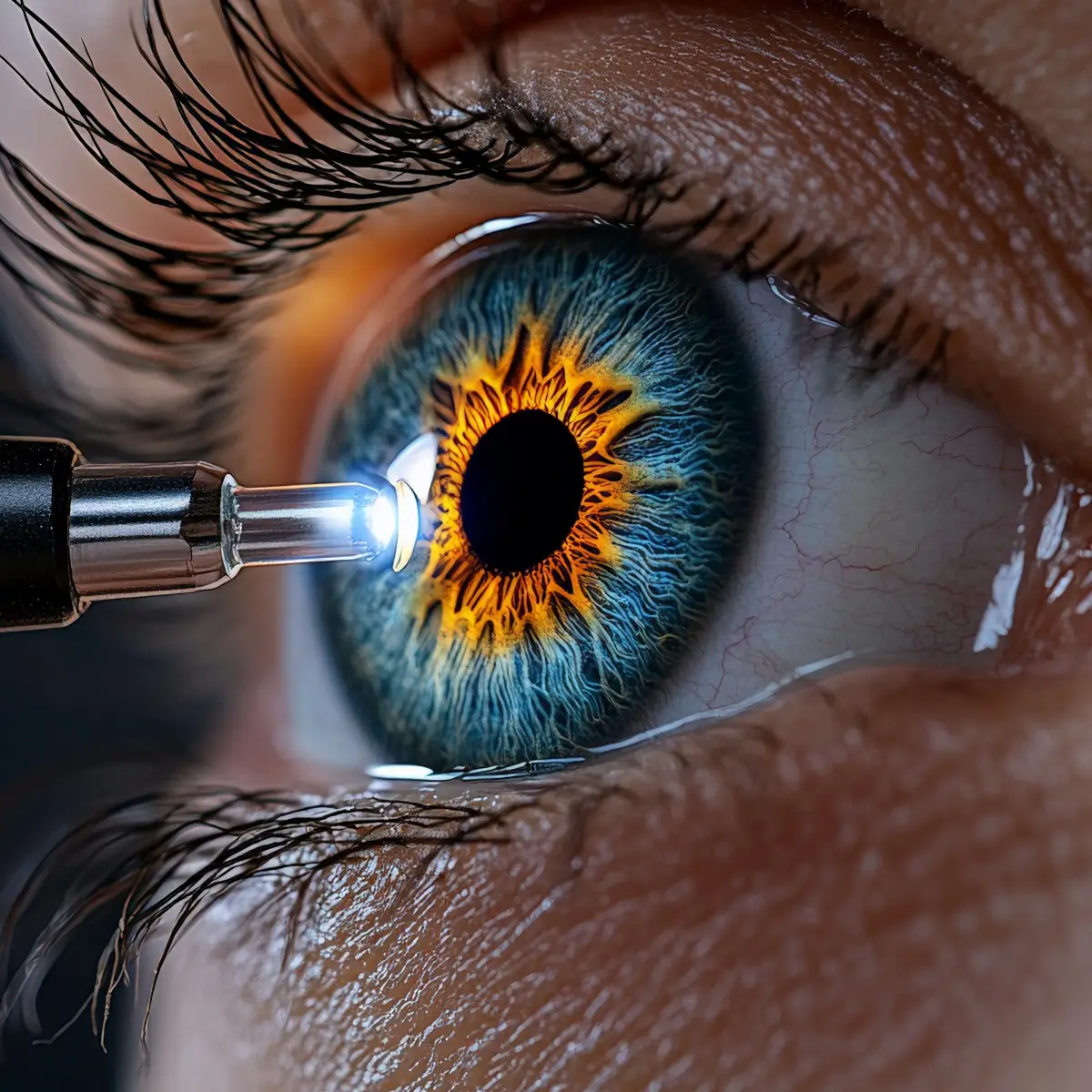
Cross-Linking and Ring Segment Treatment
Keratoconus Cross-Linking and Ring Segment Treatment: What Does It Involve?
Keratoconus
Prof. Dr. Efekan Coşkunseven discusses keratoconus on the Saglik Hattı television programme.
Keratoconus
In a health segment on Kanal 24, Prof. Dr. Efekan Coşkunseven discusses keratoconus.
Keratoconus
In a television program hosted by Ms. Derya Baykal, Prof. Dr. Efekan Coşkunseven shares information about keratoconus.
Keratoconus
Prof. Dr. Efekan Coşkunseven discusses keratoconus on CNN TÜRK's Yaşama Sevinci television programme.
Keratoconus
Prof. Dr. Efekan Coşkunseven discusses keratoconus on the Haber Türk television program.
Keratoconus
Prof. Dr. Efekan Coşkunseven discusses keratoconus on the TV8 television programme.
Keratoconus
Prof. Dr. Efekan Coşkunseven discusses keratoconus on CNN Türk.
Keratoconus
Prof. Dr. Efekan Coşkunseven discusses keratoconus on AHaber.
Keratoconus
Prof. Dr. Efekan Coşkunseven discusses keratoconus on Kanal D.
What's in this article?
- Keratoconus Treatment: An Overview by Prof. Dr. Efekan Coşkunseven
- Why Choose Prof. Dr. Efekan Coşkunseven for Keratoconus?
- Keratoconus: What Is It?
- Why Does Keratoconus Matter?
- Keratoconus: How Does the World Look Through Affected Eyes? (Vision Simulation)
- Keratoconus: Causes and Risk Factors
- Keratoconus: What Are the Symptoms?
- How Is Keratoconus Diagnosed?
- Keratoconus Symptoms: What Are the Signs? 1-Minute Keratoconus Risk Test
- When Should You See a Doctor for Keratoconus?
- The Importance of Early Examination
- An Important Note
- What Are the Treatment Options for Keratoconus?
- Who Is More Likely to Develop Keratoconus?
- Quality of Life After Keratoconus Treatment
- The Importance of Early Examination
- Stages of Keratoconus and Its Progression
- The Importance of Early Examination
- Diagnosis and Treatment Options for Keratoconus Patients in Turkey
- Keratoconus Diagnosis, Monitoring, and Current Approaches in Istanbul
- Keratoconus Treatment Costs: What Determines the Price?
- Informative Articles on Eye Health
- Keratoconus Treatment

+50,000 Keratoconus Surgeries Performed
Keratoconus Treatment Options


Keratoconus: What Is It?
Keratoconus is an eye condition in which the cornea — the clear, curved layer at the front of the eye responsible for focusing vision — gradually thins and bulges forward into a cone-like shape. This change in corneal shape makes it difficult for light to focus properly on the retina, and can cause symptoms such as blurred vision, ghosting, or double vision.
A healthy cornea is typically round and smooth. As keratoconus develops, the central or lower portion of the cornea thins and protrudes forward. This can lead to a progressive increase in astigmatism and myopia. Patients often first notice the condition through frequently changing eyeglass prescriptions or an inability to achieve clear vision even with glasses.
Keratoconus typically begins during adolescence or early adulthood and, though it varies from person to person, can progress slowly over the years. In some patients the progression is more rapid, while in others the condition remains stable for long periods. For this reason, regular eye examinations and corneal mapping tests are important for monitoring the disease.
Keratoconus is not contagious. In most cases, a combination of factors is thought to contribute — including genetic predisposition, habitual eye rubbing, and allergic eye conditions.
Why Does Keratoconus Matter?
Keratoconus is a progressive eye condition in which the cornea gradually thins and bulges forward into a cone shape, affecting the quality of vision. Because it rarely causes noticeable pain in its early stages, it often goes undetected for some time. Patients typically first present with complaints such as frequently changing eyeglass prescriptions, deteriorating night vision, or difficulty achieving adequate clarity.
The critical importance of keratoconus lies in the significant advantage that early detection offers in preserving vision quality. Today, advanced diagnostic tools such as corneal topography and tomography make it possible to identify the condition at an early stage and establish an appropriate monitoring plan. For this reason, rapidly changing prescriptions and fluctuating vision quality — particularly in younger individuals — should not be overlooked.
In most patients, keratoconus progresses slowly over the years; however, in some individuals the progression can be more rapid. Regular eye examinations, monitoring of risk factors, and timely evaluation of appropriate management options are therefore essential. Being aware of controllable factors — in particular, the habit of eye rubbing and the presence of allergic eye disease — is important, as these can influence the course of the condition.
This article has been prepared for general informational purposes about keratoconus. Diagnosis and treatment planning require an individualised assessment and must be carried out by an ophthalmology specialist.
Keratoconus: How Does the World Look Through Affected Eyes? (Vision Simulation)
Keratoconus causes the cornea to thin and bulge forward, preventing light from focusing properly on the retina. This can give rise to a range of visual complaints. Reduced vision quality is not limited to blurring alone — double vision, shadowing, and particularly nighttime light scatter are also commonly reported symptoms.
The images below are simulation examples representing the types of visual changes that may be experienced by patients with keratoconus. These simulations are not diagnostic tools; they are intended solely to help illustrate the potential visual effects of the condition. The actual visual experience may vary depending on the stage of the disease, corneal structure, and individual factors.
"This is a simulated representation of vision. It is not intended for diagnostic purposes."
01
Blurred Vision
Due to irregularities on the corneal surface, light cannot focus at a single point. This can make it particularly difficult to read text and distinguish fine details.


- A secondary ghost image appearing around the edges of objects
- Text appearing overlapped or doubled
- Shadowing that becomes particularly noticeable around headlights and street lamps.
02
Light Scattering and Halos
Halos or light scatter — particularly around headlights, street lamps, and screen glare — are commonly noticed at night. Driving after dark may become more difficult.


- Glare or halos around headlights and street lights at night
- Lights appearing scattered or blurred
- Difficulty with night vision
03
Shadowing and Double Vision
A single object may appear as multiple images or as a ghost image. This can be associated with irregular astigmatism and varies from person to person.


- A secondary image forming around the edges of objects
- Text and shapes appearing to overlap
- Symptoms that become more pronounced in bright light
Keratoconus: Causes and Risk Factors
The precise cause of keratoconus is not yet fully understood; however, genetic, environmental, and mechanical factors are thought to act together in triggering the condition. These factors affect the structural integrity of corneal tissue and, over time, can lead to thinning and progressive changes in the shape of the cornea.
Genetic predisposition is recognized as a significant factor in the development of keratoconus. Individuals with a family history of the condition may have a higher likelihood of developing it themselves. For this reason, regular eye examinations are recommended for those with a known family history.
Eye rubbing is one of the factors most frequently associated with the progression of keratoconus. In particular, frequent and forceful eye rubbing driven by itching — especially in individuals with allergic eye disease — can create mechanical stress on the cornea, contributing to the onset or worsening of the condition.
Allergic eye disease and chronic eye itching are conditions commonly seen alongside keratoconus. Prolonged itching both increases the tendency to rub the eyes and may affect the sensitivity of corneal tissue.
Beyond these, some research suggests that factors such as biomechanical weakness of corneal tissue, connective tissue structure, and oxidative stress may also play a role in the development of the disease. However, these mechanisms can vary considerably from person to person.
Keratoconus most often arises not from a single cause, but from a combination of multiple factors acting together. Understanding these risk factors — and, in particular, reducing habitual eye rubbing — therefore represents an important preventive approach that may influence the course of the condition.
01
Genetic Factors
Keratoconus may have a genetic predisposition. Individuals with a family history of keratoconus carry a higher risk of developing the condition.
For this reason, genetic factors are thought to play a significant role in the disease.
03
Environmental and Hormonal Factors
Keratoconus tends to onset during puberty and may continue to progress into early adulthood.
This suggests that hormonal changes may play a role in the development of keratoconus.
Additionally, certain environmental factors — such as prolonged exposure to UV light — may increase the risk of keratoconus.
02
Eye Rubbing Habit:
Frequently and vigorously rubbing your eyes is one of the significant factors that increase the risk of keratoconus. Eye rubbing is particularly common in individuals with allergic eye disease or chronic eye itching. Over time, this habit can cause the corneal structure to weaken and thin.
04
Association with Connective Tissue Disorders
Individuals with certain connective tissue disorders — such as Marfan syndrome and Ehlers-Danlos syndrome — carry a higher risk of developing keratoconus.
These conditions can weaken collagen structure, which in turn compromises the integrity of the cornea.
Keratoconus: What Are the Symptoms?
The symptoms of keratoconus can vary depending on the stage of the disease. In the early stages, complaints may be mild and are often mistaken for a simple change in glasses prescription. As the disease progresses, changes in vision quality can become more pronounced.
Early-stage symptoms most commonly include blurred vision and loss of clarity. Patients may describe glare around night lights, halos around headlights, and shadowing in their vision. Frequently changing glasses prescriptions or rapidly increasing astigmatism within a short period may also be early indicators of the condition.
In the more advanced stages of the disease, as the corneal shape distortion increases, fluctuations in vision quality become more noticeable. Even when looking with one eye, patients may experience double vision, shadowed or distorted letters, and reduced vision quality — particularly in low-light conditions — all of which can affect daily life.
Some patients may also report eye fatigue, headaches, and difficulty focusing on screens for extended periods. These symptoms tend to be noticed more frequently in individuals with allergy-related eye itching or a habit of rubbing their eyes.
Keratoconus does not typically cause pain, and its symptoms can develop slowly. For this reason, it is important to consult an ophthalmologist if you notice rapid prescription changes at a young age, an inability to achieve adequate vision with glasses, or a significant deterioration in night vision.
01
Blurred or distorted vision:
The irregular shape of the cornea causes light to refract at different angles onto the retina.
02
Light Sensitivity:
Seeing "halos" around bright lights at night — such as headlights or street lamps — is common.
03
Frequent Prescription Changes:
Myopia or astigmatism values may change frequently.
04
Fluctuations in Visual Quality:
Visual acuity may vary between morning and evening hours.
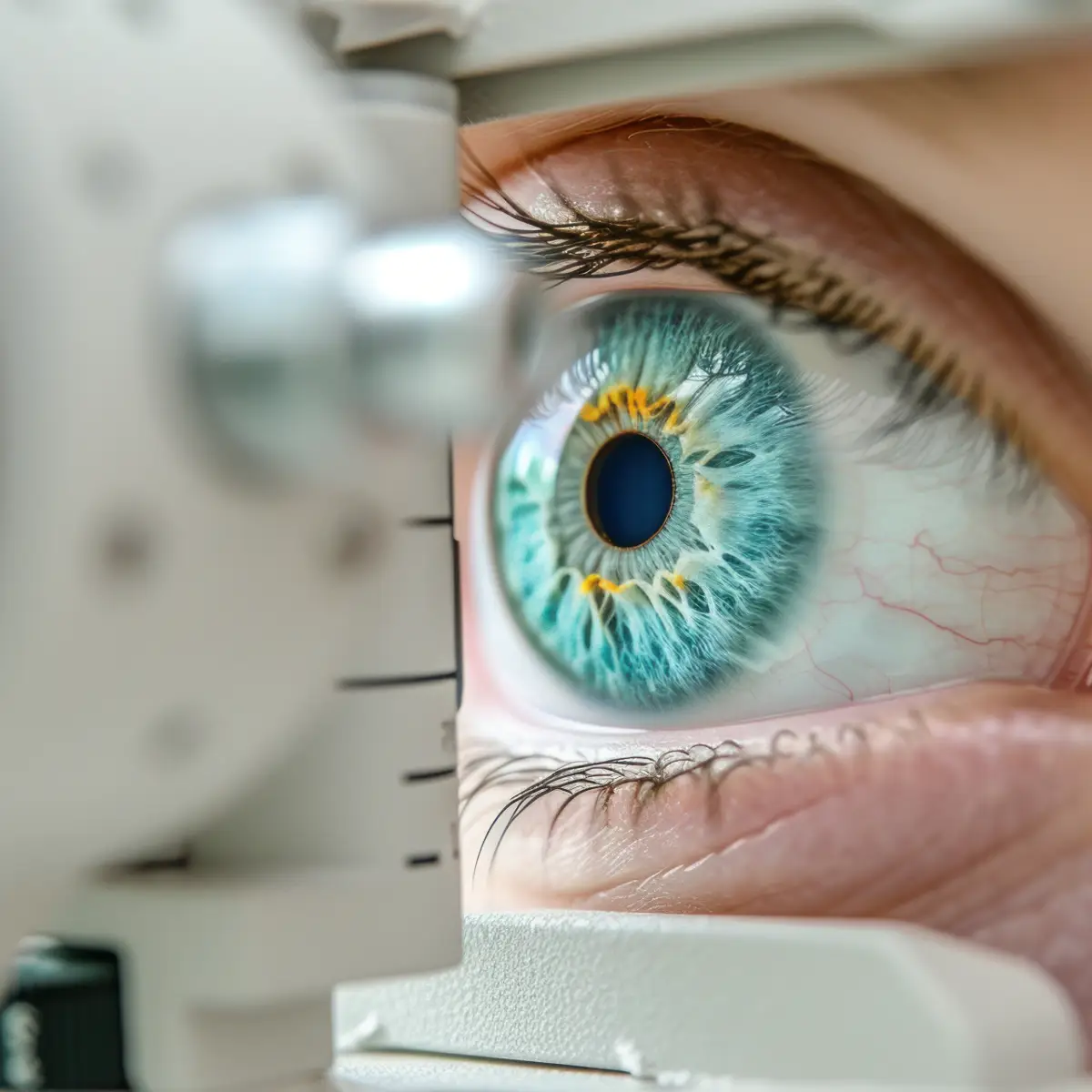
How Is Keratoconus Diagnosed?
Keratoconus is diagnosed through a detailed eye examination combined with specialized imaging techniques that evaluate the structure of the cornea. Because symptoms can be mild in the early stages of the disease, corneal analysis is essential in addition to standard vision testing.
The examination begins with measuring visual acuity and reviewing your symptoms. Rapidly changing eyeglass prescriptions, increasing astigmatism, or difficulty achieving adequate vision even with glasses can all be significant indicators of keratoconus.
One of the most important tools in diagnosing keratoconus is corneal topography — a corneal mapping test that produces a detailed map of the surface curvature of the cornea, capable of detecting even the smallest irregularities. When necessary, advanced imaging such as corneal tomography is also used to evaluate corneal thickness and the posterior corneal surface in detail.
Once a diagnosis of keratoconus has been established, regular monitoring becomes one of the most critical steps in management. The rate of progression varies from person to person, and periodic follow-up examinations allow any changes in the cornea to be tracked — ensuring that appropriate interventions can be considered in a timely manner when needed.
Keratoconus Symptoms: What Are the Signs?
1-Minute Keratoconus Risk Test
This short questionnaire provides a preliminary assessment of symptoms that may be associated with keratoconus. It does not constitute a diagnosis. A definitive evaluation requires a comprehensive eye examination and corneal topography.
Has your eyeglass prescription changed rapidly in recent years?
Do you notice glare or halos around lights at night?
Is vision blurrier in one eye compared to the other?
Do you frequently rub your eyes?
Do you experience allergic eye itching?
Has anyone in your family been diagnosed with keratoconus?
When Should You See a Doctor for Keratoconus?
Keratoconus typically progresses slowly and without pain. As a result, the earliest signs can easily be mistaken for a simple refractive problem requiring a new glasses prescription. Certain symptoms, however, may indicate that a detailed evaluation of the corneal structure is warranted.
If you are experiencing frequent prescription changes at a young age, increasing astigmatism, difficulty seeing clearly even with glasses, or pronounced glare and halos around lights at night, it is important to consult an ophthalmologist without delay. Early evaluation is critical for accurately determining the stage of the disease, assessing the risk of progression, and establishing an appropriate follow-up plan.
The Importance of Early Examination
Early diagnosis in keratoconus is essential for assessing the risk of disease progression and establishing an appropriate monitoring plan. Advanced imaging techniques now available — such as corneal topography and tomography — allow the condition to be detected even in its earliest stages.
An early evaluation can help bring the risk of progression under control and reduce the likelihood of requiring more advanced surgical interventions.
An Important Note
Because keratoconus rarely causes pain, many patients do not notice the problem until it has already advanced. For this reason, any unexplained changes in vision quality — particularly in younger individuals — warrant prompt evaluation by an ophthalmology specialist.
Regular eye examinations remain the single most important step for both early diagnosis and ongoing monitoring of the condition.
What Are the Treatment Options for Keratoconus?
The goal of keratoconus treatment is not to "completely eliminate" the disease, but to control its progression, preserve corneal structure, and help you achieve the best possible visual quality. For this reason, the approaches that may be applied can vary depending on the stage of the disease, corneal thickness, your age, visual expectations, and the demands of your daily life.
In early stages, eyeglasses or specialized contact lenses may be sufficient, while patients at risk of progression may be evaluated for methods aimed at strengthening corneal resistance. At intermediate stages, procedures that help reshape the cornea or combined treatment approaches may come into consideration. In advanced stages, where corneal tissue has thinned significantly or vision cannot be adequately corrected by other means, surgical options are evaluated.
Today, the most important principle in keratoconus treatment is individualized planning. In many patients, rather than a single method, different treatments may need to be applied at specific intervals or in combination. For this reason, regular follow-up, early diagnosis, and correct timing are the most critical components of the treatment process.
Who Is More Likely to Develop Keratoconus?
Keratoconus can occur at any age, but it most commonly develops during adolescence and early adulthood. Because the condition tends to progress slowly, early symptoms may go unnoticed for a long time. In some individuals, the risk may be higher due to genetic predisposition or environmental factors.
Keratoconus is known to occur more frequently in the following groups:
Quality of Life After Keratoconus Treatment
Living with keratoconus can be challenging, particularly in advanced stages. Daily activities may be significantly affected by visual disturbances. Tasks such as reading, using a computer, and driving can become difficult for patients with keratoconus. Quality of Life After Treatment:
Improved Visual Quality
Many patients experience clearer, higher-quality vision following treatment. Glasses, specialist contact lenses, or procedures aimed at strengthening corneal stability can help improve the level of vision. Intracorneal ring segment implantation may enhance visual quality in selected patients.
Fewer Visual Limitations
A reduction in visual irregularities can make everyday activities more manageable. Tasks such as reading, working at a computer, and driving may become easier. Some patients also notice an improvement in night vision.
Long-Term Visual Stability
In suitable candidates, treatment can help slow or halt the progression of the disease. Regular follow-up examinations are important for maintaining visual quality over time.
Psychological and Social Impact
Improvements in visual function can help patients move through daily life with greater confidence and participate more actively in social activities. This can have a meaningful positive effect on overall quality of life.
The Importance of Early Examination
Early diagnosis in keratoconus is critically important for assessing the risk of disease progression and establishing an appropriate monitoring plan. Advanced imaging technologies used today — such as corneal topography and tomography — allow the condition to be detected even in its earliest stages.
Stages of Keratoconus and Its Progression
Keratoconus is generally a slowly progressive condition, although the rate of progression varies from person to person. In some patients it may remain stable for many years, while younger individuals tend to experience more rapid progression. Regular monitoring and corneal measurements are therefore essential for assessing how the disease is evolving.
The stages of keratoconus are typically assessed based on measurements such as corneal curvature, corneal thickness, and visual acuity. In clinical practice, the condition is most commonly classified as early stage, moderate stage, and advanced stage.
Early-Stage Keratoconus
At this stage, vision can usually be corrected with glasses. Corneal topography may reveal mild irregularities, and the condition is often detected during routine examinations. Early diagnosis is critically important at this point.
Moderate-Stage Keratoconus
Corneal curvature increases and achieving clear vision with glasses may become more difficult. At this stage, specialty contact lenses or approaches aimed at controlling disease progression may be considered.
Advanced-Stage Keratoconus
Corneal thinning and irregularity become more pronounced. Vision can be significantly affected, and surgical options may become necessary for some patients.
Disease Progression and Monitoring
The rate at which keratoconus progresses varies from person to person. Regular corneal mapping and eye examinations are essential for monitoring the course of the disease.
The Importance of Early Examination
Early diagnosis in keratoconus is critical for assessing the risk of disease progression and establishing an appropriate monitoring plan. Advanced imaging technologies now in use — including corneal topography and tomography — make it possible to detect the condition even at very early stages.
Diagnosis and Treatment Options for Keratoconus Patients in Turkey
Advances in diagnostic and therapeutic technologies in ophthalmology have made it possible to detect and monitor keratoconus earlier and with greater precision. Many eye centers and university hospitals across Turkey use advanced corneal diagnostic devices and surgical techniques in the management of corneal diseases.
Advanced imaging methods such as corneal topography and tomography are widely used in the evaluation of keratoconus, enabling detection even in its earliest stages. Early diagnosis plays a critical role in assessing the risk of disease progression and establishing an appropriate monitoring plan.
Approaches used in keratoconus management in Turkey may include specialized contact lens fitting for visual rehabilitation, procedures aimed at strengthening corneal biomechanics, and surgical interventions in selected patients. The most appropriate treatment option is determined on an individual basis, taking into account the patient's corneal structure and the stage of the disease.
Turkey has also become an increasingly preferred destination for international patients seeking care in ophthalmology. Key factors behind this trend include experienced physicians, advanced technological infrastructure, and a multidisciplinary approach to care. That said, the most suitable assessment and treatment plan must be determined individually for each patient, with clinical decisions made following a thorough examination.
Advanced Diagnostic Technologies
Many centers in Turkey can detect keratoconus at an early stage using corneal topography, tomography, and pachymetry analysis.
Current Treatment Approaches
Options include visual rehabilitation, procedures aimed at strengthening corneal integrity, and surgical interventions for selected patients.
Experienced Corneal Specialists
Physicians with extensive expertise in corneal diseases, combined with modern surgical techniques, play a central role in treatment planning.
Infrastructure for International Patients
In recent years, Turkey has become an increasingly sought-after destination for international patients seeking specialized eye care.
Keratoconus Diagnosis, Monitoring, and Current Approaches in Istanbul
The diagnosis and monitoring of keratoconus requires a combination of clinical experience and advanced diagnostic technologies. Istanbul has developed significant expertise in this field, with specialized centers for eye diseases and ophthalmologists who focus specifically on corneal conditions. For this reason, keratoconus in Istanbul is a destination sought by patients from both within Turkey and abroad for diagnosis and ongoing monitoring.
The treatment approaches available for keratoconus are determined by the stage of the disease and the structural characteristics of the cornea. For patients searching for keratoconus treatment in Istanbul, the most important first step is a thorough examination and an individualized clinical assessment.
Experienced corneal specialists use advanced diagnostic methods — including corneal topography and tomography — to determine the stage of the disease and develop an appropriate monitoring plan. For patients specifically looking for a corneal specialist in Istanbul, accurate evaluation is particularly critical for conditions such as keratoconus, which require dedicated expertise.
Prof. Dr. Efekan Coşkunseven continues his work in corneal diseases and keratoconus through extensive clinical experience, scientific research, and presentations at international conferences. The diagnosis, follow-up, and individualized treatment planning for keratoconus patients are guided by modern diagnostic methods and current clinical approaches.
Because the corneal structure and disease progression can differ from one individual to the next, the assessment process for keratoconus must be conducted through a detailed examination and comprehensive diagnostic investigations.
Keratoconus Treatment Costs: What Determines the Price?
The cost of keratoconus treatment varies depending on the stage of the disease, the procedure to be performed, and the follow-up process — rather than a single fixed fee. For this reason, an accurate price can only be determined after a detailed examination and evaluation for each patient.
Keratoconus is a corneal disease that progresses differently in each patient. In some cases, glasses or specialty contact lenses may be sufficient, while others may require cross-linking, intracorneal ring segments, or a combination of approaches. These differences influence the treatment plan and, consequently, the overall cost.
The main factors that affect treatment costs include:
-
The stage of the disease and corneal structure
-
The treatment method or combination of methods to be applied
-
The technology and surgical equipment used
-
Post-procedure follow-up and monitoring
-
The patient's visual needs and expectations
While a single treatment may be sufficient in some cases, others may require more than one procedure to halt disease progression and improve visual quality. The treatment process is therefore planned on an individual basis for each patient.
For the most accurate assessment, a thorough eye examination, corneal topography, and any necessary diagnostic tests are essential. Following the examination, you will receive detailed information about the most appropriate approach and the treatment process.